One of the greatest pieces of physiological research in the twentieth century was undertaken by William Garner Sutherland D.O. He was one of the early graduates of the original American School of Osteopathy at Kirksville, Missouri in 1900. Whilst a student he noticed that the structure of certain cranial bones, particularly where they joined each other, were bevelled in a striking manner. He noted that there was a marked internal bevel where the squama of the temporal bone overlaps the great wing of the spheroid and the inferior border of the parietal, which itself displayed a marked external bevel. As he was studying under Dr Still, Sutherland was very conscious of the relationship between structure and function. If these bones were so structured, then, he reasoned, there must be a physiological function related to it. Further investigation of the bones of the skull led him to note many other articulations, such as the 'tongue and groove' junction between the lateral part of the basilar portion of the occiput, where it fits into the medial aspect of the anterior third of the petrous portion of the temporal bone.
Sutherland reasoned that these joints could only make sense if they contributed towards motion between the bones. Against all the accepted medical thinking he reasoned, studied and observed the cranial structures and their functions, with a view to establishing what these were. Gradually, over many years he came to understand the inter-relationship between the bony structures of the cranium and its contents and functions. These include not only the nerve and brain tissues but strong fibrous bands which divide and support the various areas of the brain and which are intimately involved in the motion of the cranial structures. The two main tension membranes are the Falx Cerebri and the Tentorium Cerebeli.
 |
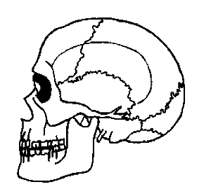
| Side View of Adult Cranium |
 |
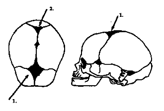
Infant Cranium Showing:
1. Anterior Fontanelle; 2. Posterior Fontanelle. |
 |
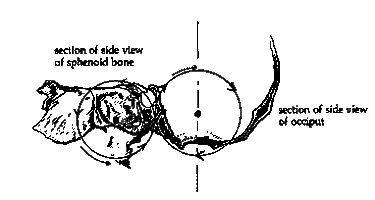
Section of side view
of speroid bone. |
Section of side view
of occiput |

|
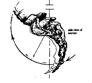
Side view of sacrum. |
| The arrows indicate directions of synchronous co-ordinated alterations in position of the main components of the cranio-sacral mechanism, during flexion (inhalation). The reverse, a return to a neutral position, occurs during extension (exhalation). The dotted line indicates the sacral position at the limit of flexion (inhalation), |
Illustration shows cranial and sacral movement during inhalation and exhalation
The erroneous belief that the skull is a rigid bony structure, and that the sutures are immovable arose from anatomists studying these structures from dried specimens. The study of living bones is quite different. It is a simple matter to feel the resilience of the skull in the living skull, even into adult life.