If the eyes are the "windows of the soul," then the mouth is the "doorway to the body. " The oral cavity is situated at the end of the digestive tract and is surrounded by the lips and cheeks externally and by the gums and teeth internally. The mouth cavity is connected to the pharynx and is covered at the top by the hard and soft palates. The tongue forms the large part of the cavity's floor. The activities of these component parts require fluid, which comes from the salivary glands.
The Jaw
It is hard to believe that a backache or a pain in the legs can originate in the jaw. However, an understanding of the relationship between the muscles and bones in the jaw and the rest of the body leads to an understanding of how an imbalance in one part affects another.
The jaw bears the teeth and forms the framework of the mouth. It consists of two bones: the maxilla (upper jaw) and mandible (lower jaw). The upper part of the jaw is stationary, while the lower part is the only movable bone in the face. The position of the jaws is dependent on the relationship of the teeth to each other when the mouth is closed.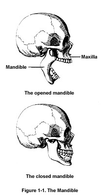
The mandible or the lower part of the jaw functions like a hinge, permitting the mouth to open and close, as shown in Figure 1.1. This hinge action is accomplished by bones and muscles located in the skull, neck, and face. There are two phases involved in opening and closing the jaw. The first phase is a simple hinge action. The second phase involves a gliding action of the joint, which helps open the jaw to its maximum. The muscles that coordinate the movements of the jaw joints originate in the head, neck, and face areas. Due to the relationship of these muscles to the muscles of the shoulder and back, any imbalance in the jaw area may eventually affect the shoulder and back.
The temporal bone of the skull (the bone on the side of the head, above the ears), and the mandible fit together to form the jaw joint called the temporomandibular joint as shown in Figure 1.2. There are two such joints-one on the left side of the head and one on the right side.
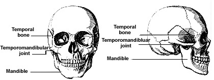
Figure 1-2. Temporal Bone and Mandible Form the Temporomandibular Joint
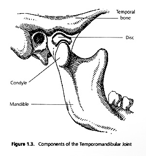 Figure 1.3 shows the components of the temporomandibular joint. Each joint is surrounded by a saclike sheath called a capsule.This capsule contains synovial fluid, which lubricates and nourishes the joints. At each end of the lower jaw, there is a knoblike structure called a condyle. At the ends of bones throughout the body, condyles are attached to muscles that join the bone to nearby bones. Mounted on top of each condyle is a cushion made of cartilage and known as the disc. As with any joint in the body, a disc or cushion sits between the joint and its socket, which, in the case of the jaw, is located on the skull. This disc moves with the condyle and prevents it from hitting the temporal bone. When the disc is abnormally placed, the jaw may not function properly causing pain or discomfort in the head, neck, shoulder, and back area. Since the jaw joint sits right in front of the ear, any problems with this joint area may also affect the ears, eyes, and sinuses.
Opening and closing the jaw is a relatively simple movement compared with the joint movement that occurs while grinding the teeth during the act of chewing. During grinding, one jaw joint slides forward, while one slides back. This movement can be felt by placing your fingers in front of your ears while making grinding movements with your teeth. If no problems exist, this motion is performed with ease, and quietly. However, audible clicking or popping noises or pain may indicate that the joints are not functioning normally. (See TMJ in Part Two.)
The Teeth
Teeth-they're not just for chewing. Our teeth perform important functions such as helping us to eat and speak, and keeping our facial muscles from sinking in. Teeth are not for opening bottles, holding nails and pins, or biting fingernails or pencils. If mistreated, teeth are subject to breakage, cavities, yellowing, and other forms of degeneration. Caring for your teeth requires that you see the mouth and teeth as important organs of your body.
Teeth are an integral and growing part of the body. The same blood supply that goes to the heart takes oxygen and nutrients to every tooth. And, as anyone who has ever had a toothache knows, the teeth are very much alive with nerves.
The better we understand the structure and development of teeth, the greater our appreciation of them will be. Practicing proper oral hygiene and maintaining good nutritional habits will help keep teeth and gums healthy. To better maintain our teeth, let's begin with a discussion of their structure and development.
Structure
The same structure can be seen in all human teeth, whether they are baby teeth (also known as deciduous or milk teeth) or permanent teeth. The care given to the baby teeth will be reflected by the adult teeth. Preventive care must begin even before the first tooth appears because teeth begin to develop before they erupt. Parents are now aware, for instance, of the damage done to teeth when infants are allowed to sleep with bottles in their mouths. (See Bottle Mouth Syndrome in Part Two.)
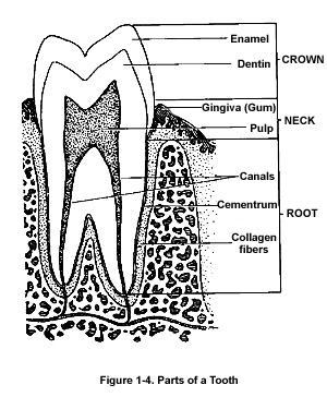 As seen in Figure 1.4, there are three main parts of a tooth-the crown, neck, and root. When you look at a tooth, the top part, called the crown, is visible. The root is the part that is imbedded in bone and covered by the gums, unless gum disease exists. The junction between the crown and the roots is called the neck. The parts of a tooth are composed of various materials.
The outer surface of the tooth, the enamel, is a hard layer covering the crown of the tooth and the upper part of the neck. The function of enamel is to resist abrasive wear and protect the tooth from damage and pain. It is the hardest tissue of the body and is composed almost entirely (97 percent) of inorganic salts. Inorganic salts, in general, are mineral constituents of the body and play specific roles in the functions of cells.
Most of the inner bulk of the tooth is made of dentin. Dentin is 67 percent inorganic salts and is not as strong as enamel. Specialized cells in dentin called odontoblasts form new dentin from minerals transported by the blood. Small tubelike structures in dentin transmit pain sensations to the nerves in the pulp, the soft tissue containing the nerve and blood supply of the tooth. When dentin is exposed by thinning or other forms of damage to enamel, the tooth may become sensitive. When tooth decay or a cavity reaches the pulp, treatment of the nerve or loss of the tooth may be inevitable.
Cementum, a bony substance, is very thin and covers the surface of the roots. Its purpose is to attach the tooth to the jawbone and the gums. When the gums are receded, cementum is exposed, causing sensitivity to hot, cold, or pressure.
The teeth rest in a bony socket. They are attached to this bone by collagen fibers, fibrous tissue that forms a bridge from the cementum to the socket. The gums or gingiva surround the teeth and the bone.
Development
When the fetus is only five weeks old, tooth buds appear. Initially, four tooth buds, representing baby teeth, appear on each side of the upper and lower jaw. These buds undergo several changes as each layer and part of the tooth begins to form. Enamel cells form and multiply. Some of these cells begin to become specialized, forming the dentin and pulp. Next, the cells for the future crown and root begin to be arranged, and layers of enamel and dentin begin to be deposited in incremental layers. The next stage is calcification or hardening of the calcium salts in the teeth, which is followed by eruption.
The process continues until approximately twenty-three weeks in utero for some teeth and up to eleven months after birth for others. Different teeth develop within this range of time; for example, front baby teeth begin to develop at five months in utero and continue until they erupt when an infant is five to seven months old. Different parts of a tooth also develop at different times. The enamel of the front baby teeth begins to develop at fourteen weeks in utero, and it is completed one and a half months after birth.
Trauma during birth, diarrhea or vomiting, feeding difficulties, rickets, and other chronic childhood diseases may cause disturbances that result in malformation of teeth. Because teeth begin to develop in utero, fetal trauma affects color or structure of teeth. Depending on when the trauma is experienced, a particular tooth or part of a tooth may be affected. For example, congenital syphilis, an infectious disease sometimes contracted during birth, will affect the teeth that are developing at that time. The front baby teeth will have notches on the edge, and the back teeth will have a pitted appearance.
Long-term treatment with the antibiotic tetracycline and excessively high amounts of fluoride in the water will cause tooth discoloration. With tetracycline, brownish bands will appear on teeth. If fluoride in excess of one part per million is ingested during the development of enamel and dentin, mottling of teeth will result. The mottling gives the teeth an opaque, chalky appearance. The severity of the condition is determined by the amount of fluoride ingested.
Eruption
There are twenty baby teeth that usually begin to appear (erupt) when a child is about six months of age. Additional teeth will then appear at the rate of about one per month. There is usually a range of plus or minus two to six months when teeth erupt and when they shed (fall out). Usually, the teeth of slender children erupt and shed earlier than the teeth of stocky children. The following table in the right column shows the approximate age at which each tooth appears.
The position of the teeth as they erupt depends on many factors. The teeth on either side and the teeth directly opposing each tooth help give guidance for proper position. If the teeth on either side or any opposing teeth are missing, a tooth may erupt incorrectly.
Age at Which Teeth Appear
|
Deciduous
Teeth |
Age
(in months) |
Permanent
Teeth |
Age
(in years) |
|
Lower central
incisors |
5-9 |
First molars |
5-7 |
|
Upper central
incisors |
8-12 |
Incisors |
6-8 |
|
Upper lateral
incisors |
10-12 |
Bicuspids |
9-12 |
|
| First molars |
10-16 |
Second molars |
11-13 |
|
| Canines |
16-20 |
Third molars |
17-25 |
|
| Second molars |
20-30 |
|
|
|
For example, if a bottom front tooth, which normally contacts the top front tooth, is lost, the top tooth will continue to erupt, since its opposing tooth is missing. If an adjacent tooth is lost, teeth on either side of the missing tooth will begin to shift into the space. This is why when a tooth is pulled, a replacement should be made to prevent shifting or extrusion of teeth. (See Tooth, Loss of, in Part Two.)